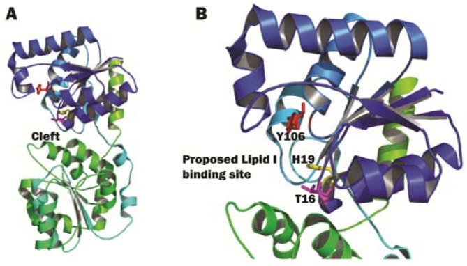
Figure 9.
E. coli MurG structure. (A) The complete view of E. coli MurG, N-domain in blue, C-domain in green. The cleft in between the two domains is indicated by an arrow; (B) A close-up view of the cleft between the N- and C-domains of MurG. Residues T16 (pink), H19 (yellow) and Y106 (red) are shown as sticks. The proposed Lipid I binding site is indicated by an arrow. The image was obtained and rendered using Pymol.

