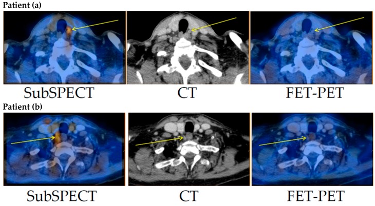
Figure 1.
Tracer-uptake in the parathyroid adenomas in patients (a) and (b). Left: Subtraction (Tc-99m-sestamibi minus I-123)-SPECT/CT. Middle: CT. Right: FET-PET/CT. Thin arrows mark the surgically confirmed location of the parathyroid adenoma. FET: O-2-(18F)fluoroethyl-l-tyrosine; PET: positron emission tomography; SPECT/CT: single photon emission computed tomography/CT.

