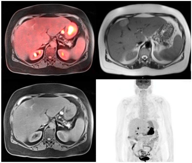
Figure 2.
64-year-old female for initial staging of gastric cancer. The PET-MRI transaxial fusion image shows intense hypermetabolic activity in the gastric cancer with SUV 29.2 and a hypermetabolic enlarged gastrohepatic lymph node (top left). T1 radial vibe transaxial image with fat suppression shows the gastric cancer to be of low to isointense signal and the metastatic lesion to be hyperintense (bottom left). T2 haste transaxial image shows a hyperintense liver lesion that was not active on the PET images which is consistent with a hemangioma (top right). The PET MIP image shows the intense activity in the stomach with activity in the adjacent gastrohepatic lymph node (bottom right).

