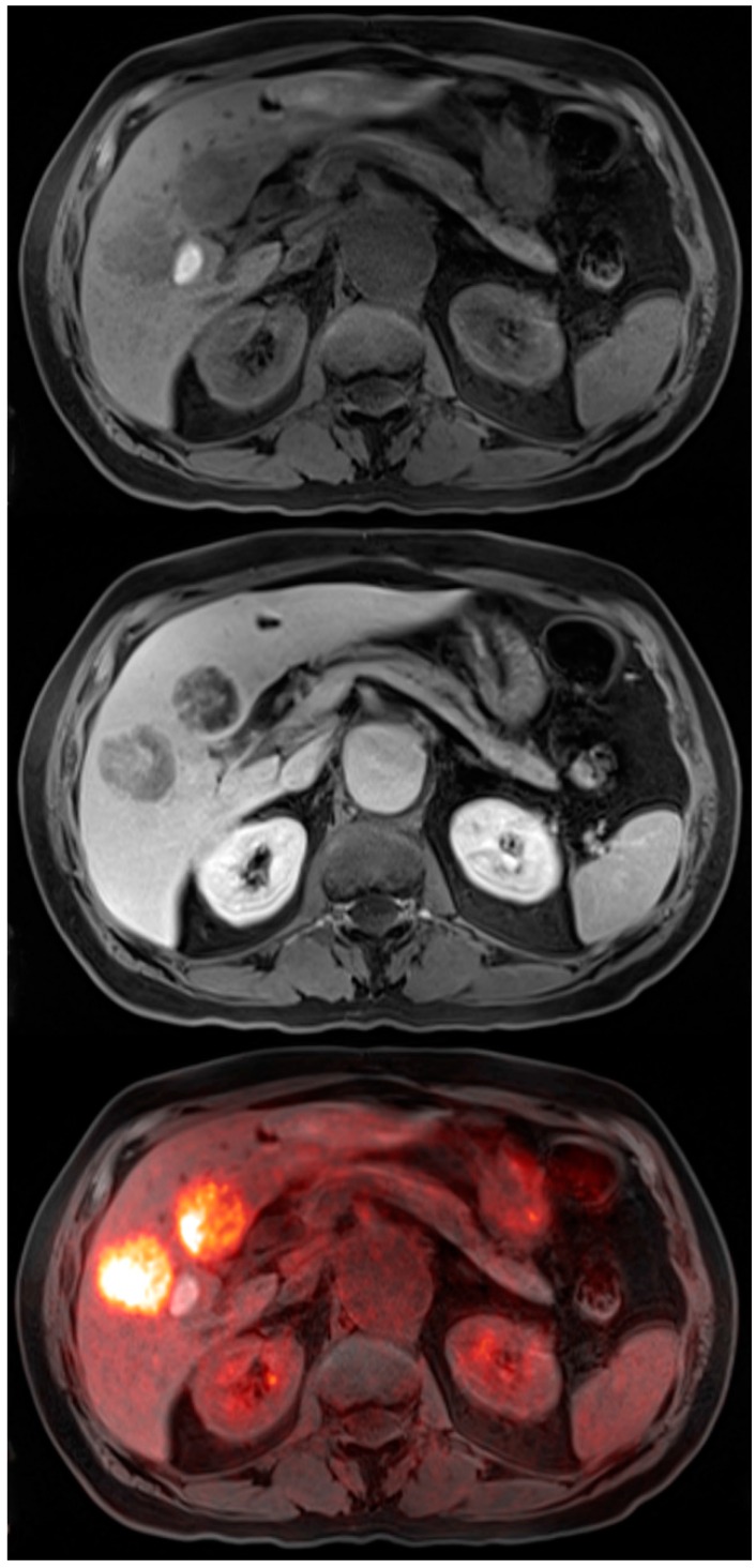
Figure 3.
72-year-old male with colon cancer. T1 precontrast transaxial image shows two low signal masses in segment 4B and 5 of the liver (top). T1 post contrast transaxial imaging with gadoxetate disodium reveals heterogenous enhancement of these two masses consistent with metastatic colon cancer (middle). PET-MRI transaxial fusion image confirms malignancy by demonstrating intense FDG activity (bottom). Also note the adjacent benign hemorrhagic cyst with high T1 signal that does not exhibit increased metabolic activity on the fusion images.

