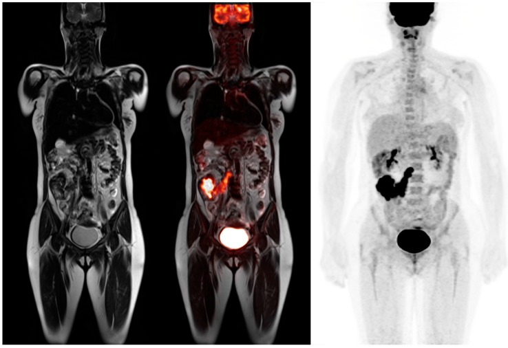
Figure 4.
45-year-old female for initial staging of colon cancer. T2 coronal image shows a large right colon mass with multiple low signal intensity lymph nodes (left). PET-MRI coronal fusion images better reveal the colon mass and chain of mesenteric lymph nodes that extends to the paracaval region (middle). PET MIP image shows the prominent FDG activity in the colon cancer and lymph node metastases (right).

