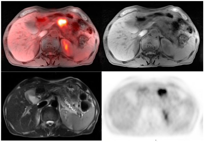
Figure 5.
62-year-old male for initial staging of pancreatic adenocarcinoma. The PET-MRI axial fusion image shows a large hypermetabolic mass in the body of the pancreas (top left). The T1 axial image demonstrates a mass in the pancreatic body that is isointense to the normal pancreatic tissue (top right). The T2 axial images show considerable main pancreatic duct dilatation (bottom left). The PET axial image displays intense metabolic activity within the pancreatic tumor relative to the liver activity with standardized uptake value of 8.7.

