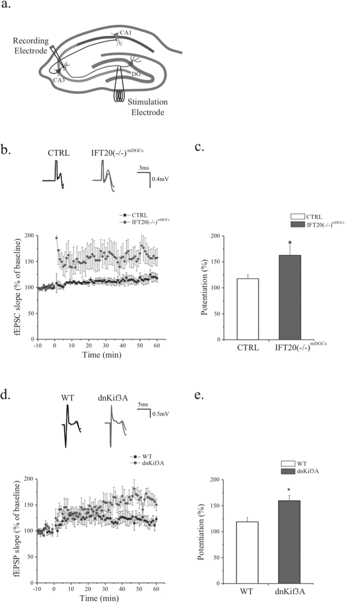
Figure 4. Removal of primary cilia from mature dentate granule cells increases synaptic plasticity in the mossy fiber pathway.
(a) A schematic representation of the tri-synaptic circuit and the position of stimulating and recording electrodes. (b) Top, representative traces of both CTRL and IFT20(−/−)mDGCs recorded before and after HFS. Bottom, IFT20(−/−)mDGCs show increased LTP compare to CTRL. (c) Average potentiation of fEPSPs slope during 50–60 minute showing enhanced LTP in IFT20(−/−)mDGCs (two-tailed unpaired t-test p = 0.048; n = 7,8). (d) Top, representative traces of both WT and dnKif3A recorded before and after HFS. Bottom, dnKif3A show increased LTP compare to WT. (e) Average potentiation of fEPSPs slope during 50–60 minute showing LTP enhancement in dnKif3A (two-tailed unpaired t-test p = 0.048; n = 5, 4). *p < 0.05; n is the number of animals.

