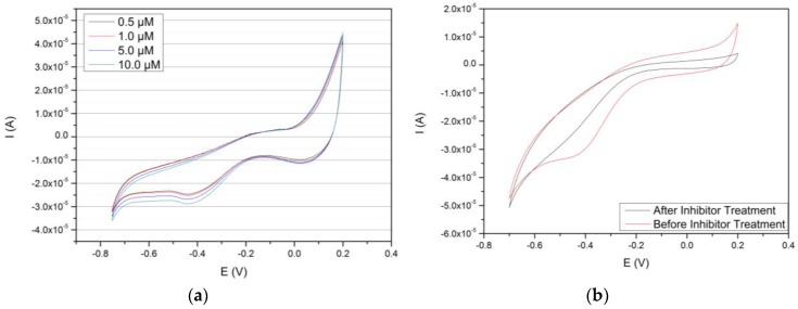
Figure 6.
Cyclic voltammograms of the Au/MPS/PAMAM G4.0-Au/CYP3A4 electrode. (a) Shows the behaviour of the electrode with 0, 0.5, 1.0, 5.0 and 10.0 µM of caffeine, respectively. An increasing, reductive current can be observed at ~ −400 mV. (b) Depicts two voltammograms produced before and after treatment with CYP3A4 inhibitor eryhtromycin and under presence of substrate caffeine. An almost total disappearance of the typical P450-related reduction peak can be observed.

