Figure 4.
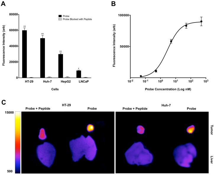
FACS study of the neoplastic cells with similar concentration of GE-137 probe showed high levels of mean fluorescent intensity and therefore high relative receptor density on HT-29 > Huh-7 > HepG2 with minimal signal on the control LNCaP cells. Competitive binding study was performed with addition of blocking peptide, which resulted in significant decrease of mean fluorescence signal intensity in all cell lines. (*: p < 0.05, **: p < 0.001) (A). GE-137 specifically binds MET receptor with Kd value of 2.9 ± 0.36 nM (B). In vivo optical imaging of the mice with subcutaneous tumors showed significant decrease in mean fluorescence signal intensity and target to background ratio 4 hours after injection of the probe and blocking peptide mixture compared to the mice that only received the probe. TBR decreased from 13.5 ± 1.2 to 1.7 ± 0.3 (p < 0.05) in HT-29 and from 5.3 ± 0.8 to 1.4 ± 0.2 (p < 0.05) in Huh-7 xenografts (C).

