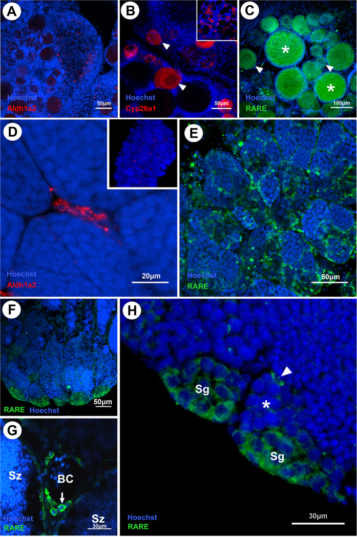
Figure 4. Fluorescent in situ hybridization of aldh1a2, cyp26a1 expression and RA responsiveness in adult gonads of medaka.
(A) aldh1a2 expression in the parenchyma cells. (B) cyp26a1 expression restricted to early oocytes; strong signal in the prophase I-arrested oocytes (arrowhead). (C) Responsiveness to RA present in early oocytes. Note the strong signal in the vitellogenic oocytes (asterisks) (D) aldh1a2 expression in the Leydig cells. (E) Responsiveness to RA present in Sertoli and Leydig cells; no germ cells appear to be responsive to RA. (F) Responsiveness to retinoic acid at the tip of the germinal epithelium lobe. (G) Leydig cells (arrow) close to blood vessels are positive for RARE driven GFP. (H) Strong GFP signal in Sertoli cells (arrowhead) and in spermatogonia closer to the tip of the germinal epithelium lobe; Small group of spermatogonia with weak expression (asterisk) more distant from the lobe. BC, blood cells; Sg, spermatogonia; Sz, spermatozoa.

