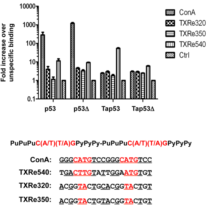
Figure 3. In vitro real-time PCR assay showing binding of indicated p53s to consensus (ConA) and three putative Trichoplax p53 response elements (TXRe540, TXRe320, TXRe350).

Binding expressed as fold increase over binding to non-specific control DNA plotted on logarithmic scale. n = 2 ± SD. Shown also is consensus p53 motif comprising two 10bp half-sites separated by up to 13 nucleotides. Nucleotides corresponding to consensus are underlined and in red.
