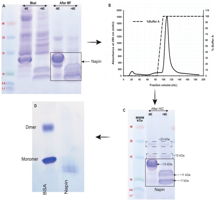
Figure 6.
Purification of napin from B. napus meal protein extract at pH 3. (A) Polypeptide profiles of meal and pH 3 extract after membrane filtration (MF); (B) Chromatograms of membrane-separated proteins further cleaned using HiTrap Phenyl Sepharose™ 6 Fast Flow Hydrophobic Interaction column (HIC); (C) Polypeptide profiles of napin after HIC; and (D) Native-PAGE separation of napin in (C). For SDS-PAGE, polypeptide profiles were under non-reducing (−ME) and reducing (+ME) conditions. Precast homogeneous gels of 20% and low range molecular weight markers (MWM) were used.

