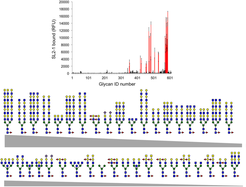
Figure 3. Carbohydrate binding specificity of SL2-1 using a mammalian glycan array.
Fluorescent dye-labeled SL2-1 (200 μg/mL) was used for glycan array screening at the Protein-Glycan Interaction Core H of the Consortium for Functional Glycomics (Emory University, Atlanta, GA). The specificity of SL2-1 was determined by testing its ability to bind to a printed array (version 5.2) consisting of 609 mammalian glycans. Of these, 40 structures contained core α1,6 fucose (upper panel, shown in red). The structures of bound core α1,6 fucosylated N-glycans are shown in decreasing order of ligand binding efficiency (see Supporting Table S2 for more details).

