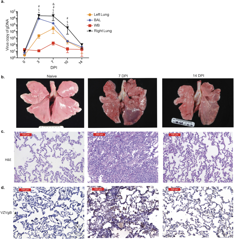
Figure 1. SVV infection results in inflammation.
(a) SVV viral loads in lung biopsies, bronchial alveolar lavage (BAL) and blood (WB) were measured by quantitative PCR using primers and probes specific for SVV ORF21 (BAL: n = 14 (0 days post infection, DPI), n = 11 (3 DPI), n = 8 (7 DPI), n = 5 (10 DPI), n = 3 (14 DPI); Lung: n = 3 (0 DPI), n = 3 (3 DPI), n = 3 (7 DPI), n = 2 (10 DPI), n = 3 (14 DPI); WB: n = 14 (0 DPI), n = 11 (3 DPI), n = 8 (7 DPI), n = 5 (10 DPI), n = 3 (14 DPI)) (#p < 0.05 for left lung; $p < 0.05 for BAL; &p < 0.05 for WB; *p < 0.05 for right lung relative to day 0). (b) SVV infection results in focal hemorrhage during peak viral replication that largely resolved 14 DPI. (c) H&E staining shows immune infiltrates and lung consolidation during peak viral replication. (d) VZVgB staining showing high levels of viral antigen 7 DPI that were decreased 14 DPI.

