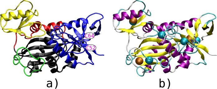
Figure 1.
(a) Three-dimensional (3D) view of the X-ray crystallographic structure of VanA, colored according to its domains: the N-terminal [A2–G121] shown in blue, the C-terminal [G212–A342] shown in black, which includes the ω-loop [L236–A256] shown in green, and the central domain [C122–S211] shown in red, which includes the opposite domain [A149–Q208] shown in yellow. The disulfide bridge C52–C64, located in the N-terminal domain, is shown with magenta labels (bottom right). (b) Localization of the collective variables (CV) used for the different TAMD calculations on a cartoon view of VanA extracted at the end of a 10 ns MD trajectory. The three structural CV are shown in orange and the five CV obtained from contact communities calculations are shown in cyan.

