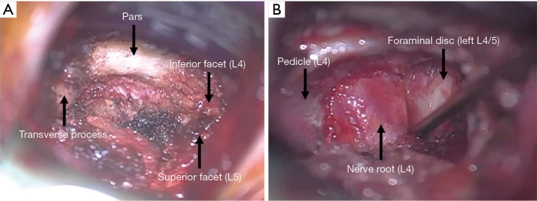
Figure 3.
Intraoperative anatomy under the microscope. (A) After the initial paramedian incision is made, the anatomy is identified; (B) after haemostasis is achieved, and the ligamentum flavum and facet joint capsule are cleared from the lateral aspect of the foramen, the nerve-disc interface can be clearly identified.

