
Fig. 1.
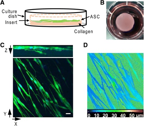
Generation of ASC sheet. a Schematic illustration of ASC sheet preparation. b Representative image of an ASC sheet in a well of a six-well plate. c 3-D projection of an ASC sheet, revealing the GFP-expressing ASCs (green) in the collagen matrix. Cell nuclei were counterstained with bisBenzimide H 33258 (blue). Bar = 20 μm. d Depth-coded image of (c). Blue and red color scale indicates the closest and furthest, respectively, from the surface of the ASC sheet. ASC adipose-derived mesenchymal stromal cell