
Fig. 7.
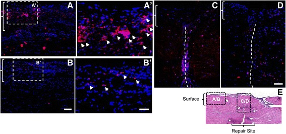
Impact of ASC sheets on tendon cell apoptosis in FDP tendons 7 days after repair. Representative immunofluorescence images of FDP tendon sections from the + HA (A, A′, and C) or + ASC sheet (B, B′, and D) groups at the tendon surface (A, A′, B, and B′) or repair site (C and D). All sections were stained for cleaved caspase 3 (red) and DAPI nuclear stain (blue). E Location index indicating the location of A–D relative to tendon repair in a tendon section with H&E staining. A–D Braces indicate epitenon regions. C, D Dotted lines indicate site of tendon transection. Bar = 100 μm for A and B, bar = 50 μm for A′ and B′, and bar = 100 μm for C and D