Fig. 1.

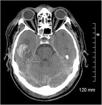
CT scan at admission. Initial CT scan taken at admission, showing right temporal parenchymal hematoma, diffuse subarachnoid haemorrhage in the left hemisphere, and diffuse axonal injury

CT scan at admission. Initial CT scan taken at admission, showing right temporal parenchymal hematoma, diffuse subarachnoid haemorrhage in the left hemisphere, and diffuse axonal injury