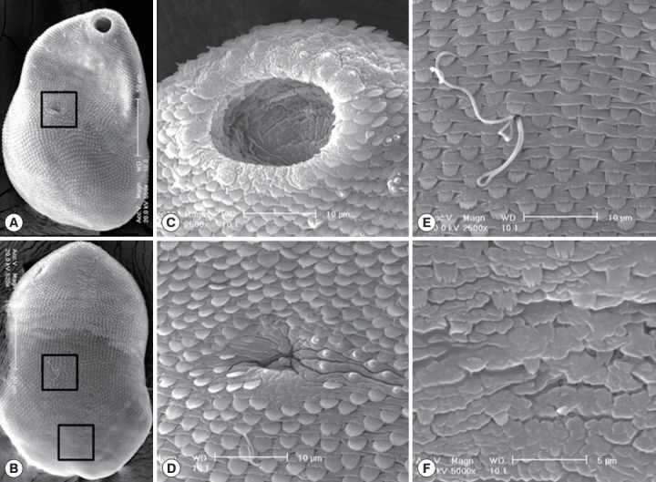
Fig. 3.

SEM findings of S. falcatus recovered from an experimental hamster. (A) Whole ventral view, showing its concave body with scale-like tegumental spines and 2 suckers, the oral sucker in anterior end and the ventral sucker dextrally located (in the square). (B) Whole dorsal view. The body surface is covered with scale-like tegumental spines except for the surface near posterior end. (C) Tegument around the oral sucker. Numerous small ciliated type I sensory papillae are seen on the dorsal lip and 2-4 grouped type I sensory papillae are observed near the oral sucker. (D) Tegument around the ventral sucker. Ventral sucker is small, dextrally located, and has 8 type I sensory papillae in it’s left margin. (E) Tegument on the dorso-middle surface. Numerous broom brush-shaped tegumental spines are compactly distributed and sperms entering into the opening of Laurer’s canal. (F) Tegument on the dorso-posterior surface. Tegumental spines here became sparser and less digitated.
