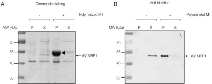
Fig. 2.

In vitro MT-binding assays using rGlMBP1. Ten µg of rGlMBP1 was incubated without or with taxol-stabilized bovine MTs (20 µM), divided into pellet (P) and soluble (S) fractions by ultracentrifugation, and then separated by 12% SDS-PAGE. (A) A SDS-PAGE gel stained with Coomassie brilliant blue. (B) Western blot using anti-histidine antibodies (1:5,000 dilution). An arrowhead (about 55 kDa) indicates MTs, whereas arrows denote rGlMTBP.
