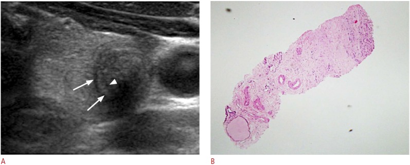
Fig. 1. A 61-year-old man with a degenerating benign nodule.
A. Transverse sonogram shows a 1.2-cm, hypoechoic solid nodule with an inner isoechoic rim (arrowhead) and a peripheral, low-echoic halo (arrows) in the left thyroid lobe. Subsequent core needle biopsy (CNB) confirmed it as degenerating nodular hyperplasia. B. A CNB specimen shows tissue consisting of a central portion of severe fibrosis and a periphery of a few follicular cells at the corresponding areas seen on sonogram (H&E, ×40).

