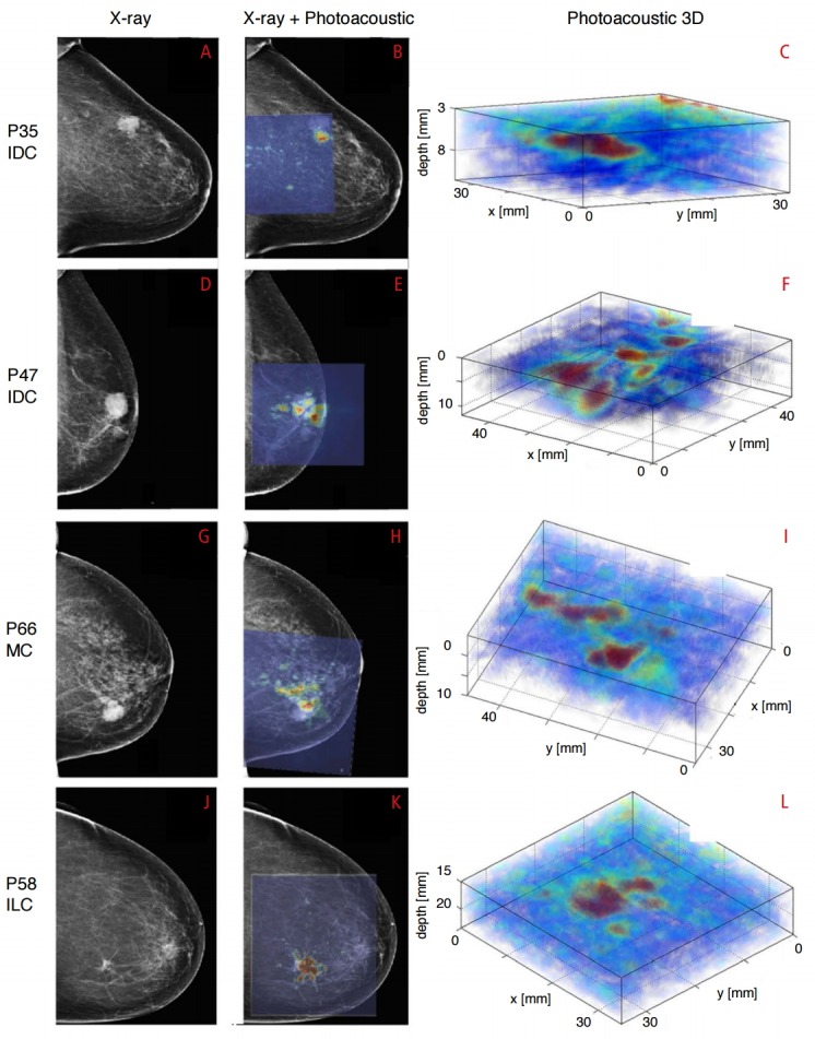
Fig. 3. Breast imaging with photoacoustic mammoscope.

Photoacoustic images were overlaid on X-ray mammograms to show lesions detected on both modalities. Reconstructed 3D photoacoustic volume encompassing each lesion of interest is also shown here. Infiltrating ductal carcinoma (IDC) lesion was seen on X-ray and photoacoustic imaging in a 79-year-old patient (A-C) and a 69-year-old patient (D-F), respectively. Mucinous carcinoma (MC) was detected in an 83-year-old patient (G-I) while infiltrating lobular carcinoma (ILC) was seen in a 65-year-old patient (J-L). The lesions were co-localized on photoacoustic images with respect to X-ray mammograms and were visualized at depths of more than 20 mm with good contrast on photoacoustic images. PXX indicates patient-identifier in the study. Reproduced from Heijblom M et al. Eur Radiol 2016, http://dx.doi.org/10.1007/s00330-016-4240-7 [55], according to the Creative Commons license.
