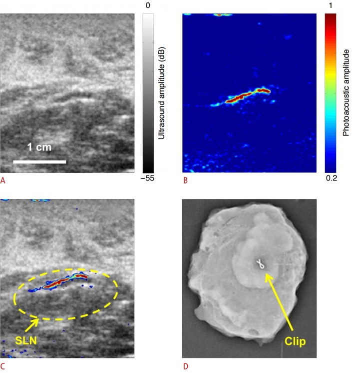
Fig. 5. Identification of sentinel lymph node (SLN) in breast cancer patients using photoacoustic imaging.

A. Sonogram shows lymph node in a breast cancer patient. B. Photoacoustic image shows a strong signal from the lymph node due to methylene blue accumulation confirming the ultrasound detected node as a SLN. C. Co-registered photoacoustic image of the SLN overlaid on sonogram. D. Radiograph of surgically removed SLN shows the presence of tissue-marking titanium clip which was implanted under the guidance of photoacoustic imaging, validating the feasibility of photoacoustic imaging to identify SLNs noninvasively. Reproduced from Garcia-Uribe A et al. Sci Rep 2015;5:15748 [75], according to the Creative Commons license.
