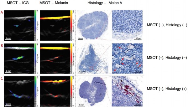
Fig. 6. Sentinel lymph node (SLN) visualization using a handheld multispectral optoacoustic tomography (MSOT) transducer in patients with melanoma.
Indocyanine green (ICG) dye was injected peritumorally to visualize the lymphatic drainage using photoacoustic imaging. While ICG indicates the location of SLNs, melanin inside the SLN suggests melanoma metastasis. A. Absence of melanin signal in ICG-positive right axillary node suggests that the patient has no detectable metastasis, confirmed as such by histology. B. Photoacoustic imaging of left axillary node (ICG-positive) in a patient suggests metastasis as indicated by strong melanin signal. Immunohistochemistry revealed no evidence of metastasis when stained for Melan A indicating false-positive photoacoustic imaging diagnosis. Left axillary lymph node in a patient showing high melanin signal on photoacoustic image (C) which was confirmed to be metastatic on histology (true positive finding). Adapted from Stoffels I et al. Sci Transl Med 2015;7:317ra199 [71], with permission of The American Association for the Advancement of Science.

