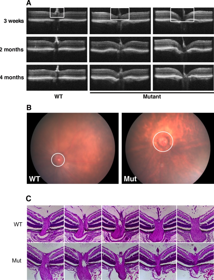
Figure 3.
Defects of optic nerve head were identified in Pitx2+/− mice. (A) Eyes from WT and mutant mice were imaged by SD-OCT for morphology of the optic nerve head at the ages indicated. White rectangle indicates the region of optic nerve head, where it is flat at all time points in wild type, but cupped with varying severity in the mutants. (B) Fundus photography at 2 months also showed morphology of optic nerve head (circle) changed in the mutant eyes. (C) Representative examples of histologic sections taken through nerve heads from a Pitx2+/+ and a Pitx2+/− eye at postnatal day 7, illustrating the absence of cupping in the heterozygous eyes.

