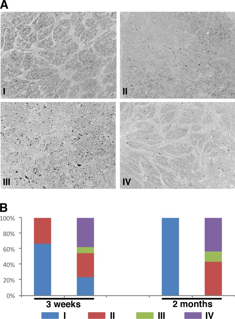
Figure 4.

Optic nerve degeneration in Pitx2+/− mice was identified by PPD staining. (A) Representative images of optic nerves from WT and mutant eyes at 2-months old following staining with PPD to visualize the myelin sheath. Optic nerves were assigned to one of four categories based on the severity of degeneration identified: (1) the majority of axons were healthy, (2) partial degeneration was identified regionally, (3) no healthy axons were seen but dying axons were still identifiable, and (4) all axons, including dying ones, were missing. Each type was indicated by a representative image. (B) Samples from 3-week-old (WT N = 12; mutant N = 13) and 2-month-old (WT N = 8; mutant N = 14) optic nerves were scored based on the severity assignment described previously. At 2 months old, all optic nerves from wild type were healthy and assigned to type I. Degeneration was found in all optic nerves from the mutants with vary severity at 2 months of age, even the density of retinal ganglion cells was normal in the corresponding retinas.
