Figure 5.
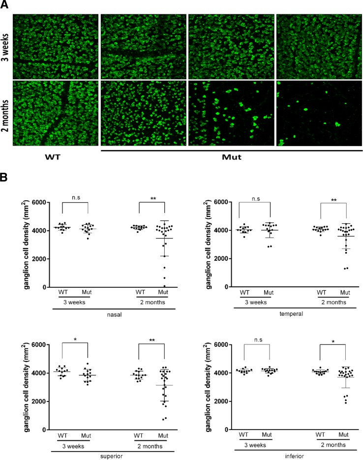
Retinal ganglion cell density is reduced in Pitx2+/− mice. (A) Flat-mounted retinas from 3 weeks, 2, and 4 months were stained with anti-RBPMS antibody, which can recognize specifically RGCs. Representative images from 3-weeks and 2-months old were presented to show fewer retinal ganglion cells in Pitx2+/− eyes. At the age of 3 weeks, there was no visible changes in RGC density. However, at the age of 2 months, RGC loss was readily seen with vary severity. (B) Retinal ganglion cell density data were analyzed by dot plotting with mean value plus ± SD. Each retina was divided into four areas: nasal, temporal, superior, and inferior. Significance: * P ≤ 0.05; ** P ≤ 0.01.

