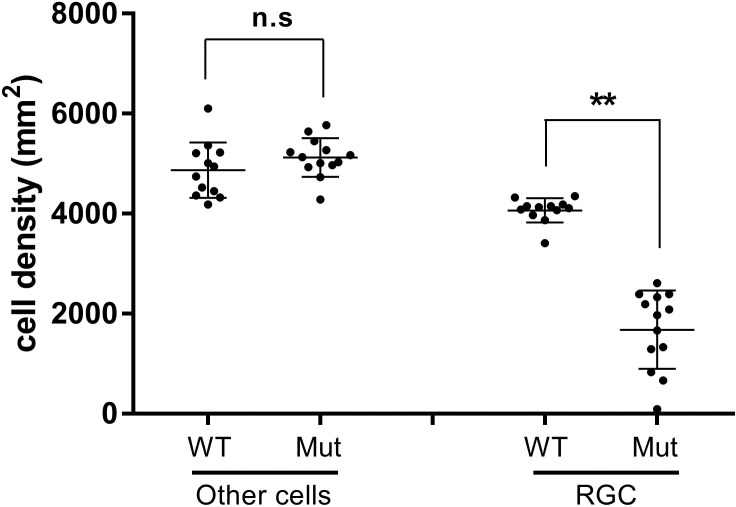
Figure 6.
Displaced amacrine cell density is unaffected in PItx2+/− mice. Comparison of Nonganglion cell densities (other cells) to ganglion cell densities in selected Pitx2+/+ versus Pitx2+/− retinas. Images (N = 13) from Pitx2+/− retinas exhibiting a ≥35% ganglion cell loss of were identified and the density of nonganglion cells “other cells” was deduced by determining the total number of DAPI+ nuclei and subtracting the number of RBPMS+ cells. “Other cells” densities from control images (N = 12) were determined in parallel. “Other cells” are presumed to represent displace amacrine cells, as they constitute the vast majority of nonganglion cells in the ganglion cell layer. Data are presented by dot plot with mean values ± SD. Significance: n.s., not significant; ** P ≤ 0.01.

