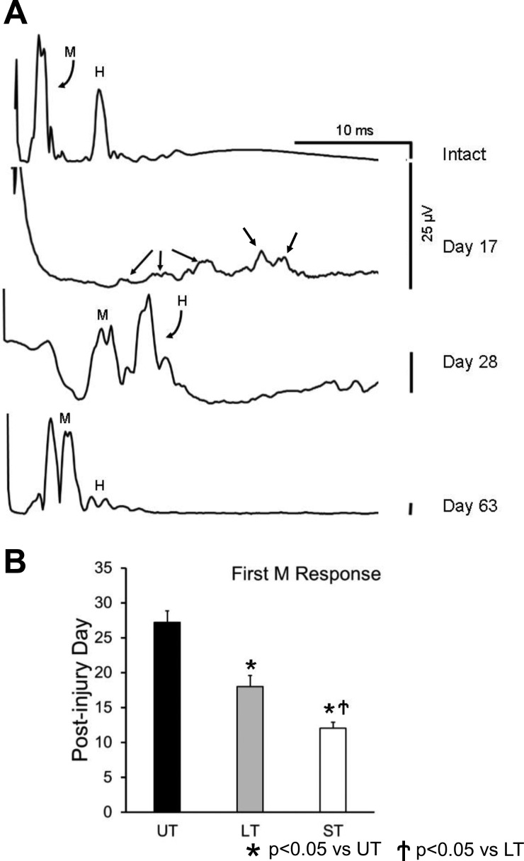
Fig. 2.
A: examples of evoked EMG responses recorded from soleus muscle of a rat in response to stimulation of the tibial nerve before (intact) and at different times after transection and surgical repair of the sciatic nerve proximal to the stimulation site are shown. Potentials designated as direct muscle (M) responses and monosynaptic H reflexes are indicated. Except for the earliest posttransection time shown (17 days), all traces are from recordings made at a stimulus intensity at which the largest H reflex was evoked, so that the posttransection timing of restoration of this reflex can be observed. At all of these postinjury times, larger M responses than shown here can be evoked at larger stimulus intensities. Each trace represents the average of at least 100 stimulus presentations. This animal was treated with two wk of daily exercise on an upslope inclined treadmill. Before day 17 after sciatic nerve transection and repair, no significant evoked soleus EMG activity could be recorded from this rat. At this earliest posttransection time that an EMG response could be evoked in this rat, multiple small potentials are observed (day 17, small arrows), making differentiation of these responses as M or H impossible. Scale bars for each of the traces are the same: 25 μV. B: mean posttransection days (±SE) when a direct muscle (M) response first could be evoked are shown for untreated (UT) rats and rats treated with exercise on a level (LT) or upslope inclined (ST) treadmill (n = 5 for UT, n = 7 for LT, n = 8 for ST).

