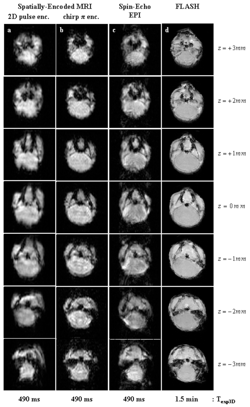
Figure 4.
Multi-slice 2D images produced by the sequences in Figure 1, recorded post-mortem on a fixated mouse. Seven 1 mm slices were acquired on the animal’s brain using a zero inter-slice gap over 3D FOVs of 16x16x7 mm3. Left to right columns: (a) SPEN MRI using a 2D slice-selective chirped excitation pulse (sequence in Fig. 1a) using the following parameters: TR=70 ms; TE=[7…22] ms; matrix size=50×50×7; pixel size=0.40×0.40×1 mm3. (b) Idem but using a chirped π-encoding pulse (sequence in Fig. 1b) and the following parameters: TR=70 ms; TE=[7…22] ms; matrix size=50×50×7; pixel size=0.40×0.40×1 mm3. (c) Spin-Echo EPI (sequence in Fig. 1c) using the following parameters: TR=70 ms; TE=15 ms; matrix size=50×50×7; pixel size=0.40×0.40×1 mm3. (d) Reference FLASH images (pixel size = 0.20×0.20×1 mm3).

