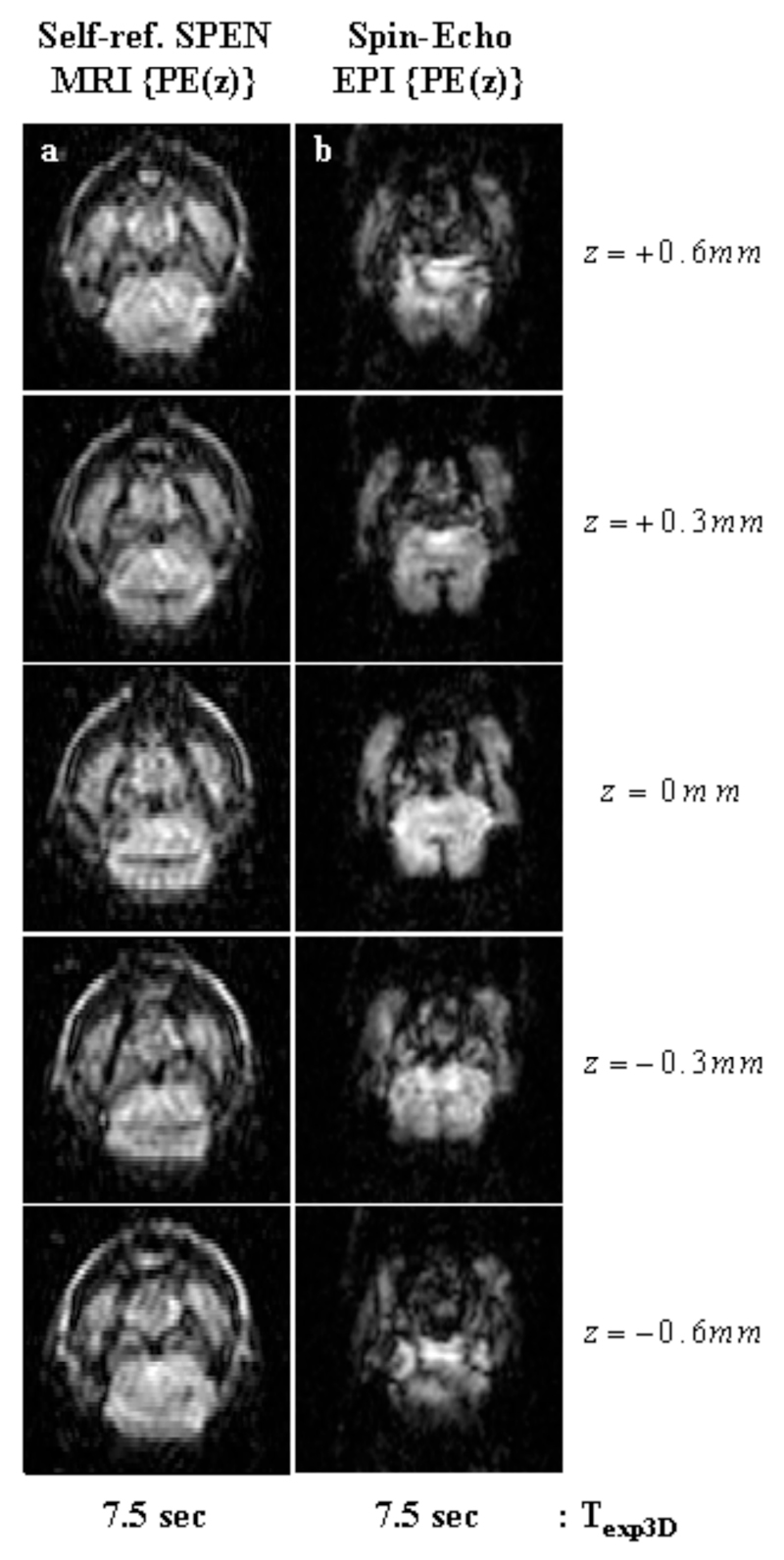
Figure 8.
In vivo 3D images of a mouse brain, using the z-axis phase-encoding sequences in Fig. 2 and leading to five slices spread over FOVs of 16x16x1.5 mm3. (a) Self-refocused phase-encoded SPEN MRI with the following parameters: TR=1.5 sec; TE=[7…25] ms; matrix size=50×45×5; pixel size = 0.40×0.45×0.30 mm3. (b) Phase-Encoded Spin-Echo EPI with the following parameters: TR=1.5 sec; TE=16; matrix size=50×45×5; pixel size=0.40×0.45×0.30 mm3.

