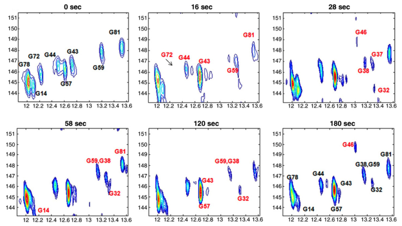
Figure 8.
Spectra from Ref. (83) illustrating the dynamic capabilities of the ultraSOFAST 2D experiment. Conformational transitions of a riboswitch are followed by recording real-time 2D 1H-15N HMQC NMR spectra. The spectra shown were recorded at pH = 6.1 and 298 K on a ∼1.7 mM 15N-G-labeled adenine riboswitch ligand-binding domain; the times indicated in each frame correspond to the time point following addition of adenine and Mg2+ to the free RNA solution.

