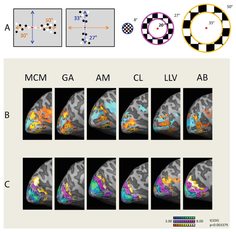
Fig. 3.
(A) The two types of stimuli used to delineate visual areas. Note that all stimuli were black and white; the color coding is for clarity. (B, C) Medial views of the left hemispheres of six participants: (B) Responses to stimulation along the verticalmeridian, in blue and cyan, and responses to stimulation along the horizontal meridian, in orange and yellow. (C) Responses to stimuli at three different eccentricities; foveal is shown in blue and green, near eccentricity in purple, and far eccentricity in orange and yellow

