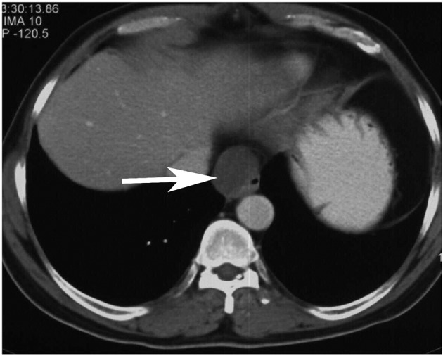
Figure 2.

Contrast-enhanced computed tomography image of the thorax shows a 3.5 × 2.3 × 3 cm well-defined homogenous cystic lesion (white arrow) along the right antero-lateral aspect of the distal esophagus focally indenting and distorting the lumen.
