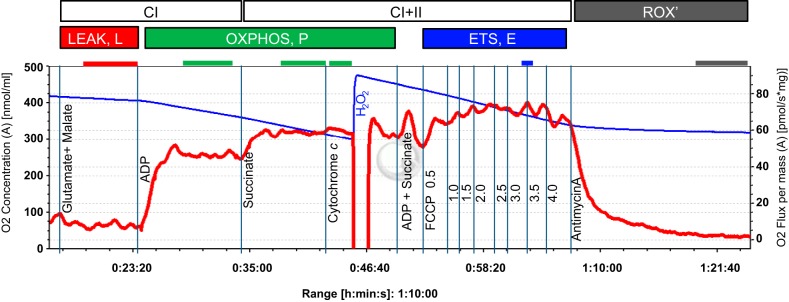
Fig. 1.
Respirometric protocol with permeabilized fibers from American Quarter Horse gluteus medius muscle. Shown is a typical trace of oxygen consumption after permeabilized fiber preparation with glutamate/malate and succinate substrate combinations to support electron flow through complex I (CI) and complex II (CII), respectively, of the mitochondrial electron transport system (ETS), and its activation by ADP. Cytochrome c was added as a quality control (see text for details), FCCP to induce uncoupling and evaluate ETS capacity, and antimycin A (inhibitor of complex III of the ETS) to evaluate residual oxygen consumption (ROX). The blue line represents the oxygen concentration (nmol/ml), the red line the muscle mass-specific O2 flux (pmol O2·s−1·mg wet wt−1; negative slope of the blue line normalized to tissue weight). Marked sections correspond to steady-state fluxes at different coupling states (L, P, and E; see text for explanations).

