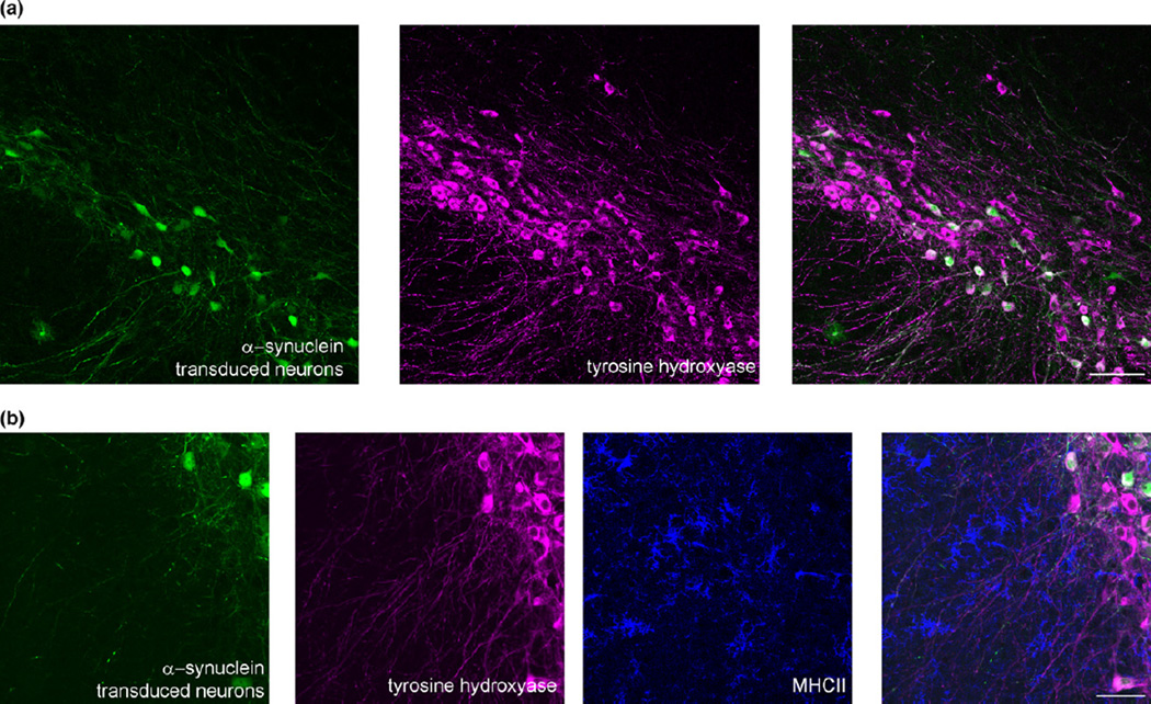
Fig. 1.
Representative images of a mouse substantia nigra pars compacta transduced with rAAV2-CBΑ-synuclein-IRES-EGFP-WPRE. (a) The eGFP shows neurons transduced with rAAV2-α-synuclein and the magenta image shows tyrosine hydroxylase-positive neurons. Scale bar = 100 µm. B) The eGFP shows neurons transduced with rAAV2-α-synuclein, the magenta image shows tyrosine hydroxylase-positive neurons, and the blue shows major histocompatibility complex II (MHCII)-positive activated microglia near α-synuclein expressing neurons. Unpublished data. Scale bar = 50 µm.

