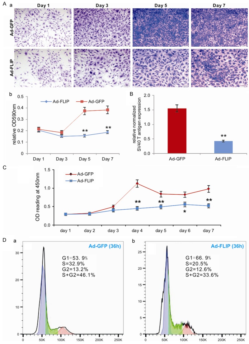
Figure 3.

The immortalization phenotype of the iMADs can be reversed by FLP recombinase. (A) Subconfluent iMADs were infected with Ad-FLP or Ad-GFP and fixed for Crystal violet staining at different time points (a). The stained cells were dissolved and quantitatively determined at A590nm (b). “**”, p<0.01 when compared between Ad-FLP and Ad-GFP infected groups. (B) Quantitative analysis of the FLP-mediated removal of SV40 T antigen in the iMADs. Subconfluent iMADs were infected with Ad-FLP or Ad-GFP. At 3 days after infection, total RNA was isolated and subjected to RT-PCR and subsequently TqPCR analysis of SV40 T antigen expression. Gapdh was used as a reference gene. “**”, p<0.01 when compared between Ad-FLP and Ad-GFP infected groups. (C) WST-1 proliferation assay. Subconfluent iMADs were infected with Ad-FLP or Ad-GFP. WST-1 substrate was added to the cell culture and assessed for A450nm readings at the indicated time points. Assays were done in triplicate. “**”, p<0.01. (D) Cell cycle analysis. Subconfluent iMADs were infected with Ad-GFP (a) or Ad-FLP (b). At 36 h after infection, cells were collected, fixed, stained with Hoechst 33342, and subjected to FACS analysis. Assays were done in triplicate, and representative results are shown.
