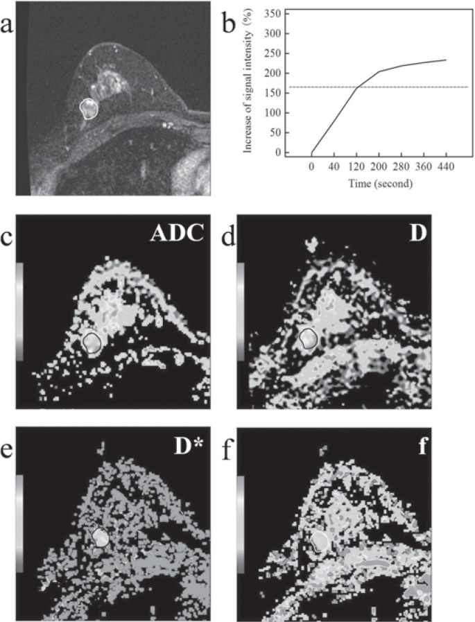
Fig. 1.
Fibroadenoma on magnetic resonance images in a 47-year-old woman. a Contrast-enhanced magnetic resonance imaging. b Time-signal intensity curve (TIC) Type I. c On diffusion-weighted images, the mean apparent diffusion coefficient (ADC) was 1.12 × 10⁻³ mm2/s. d, e, f On intravoxel incoherent motion (IVIM) images, tissue diffusivity (D), pseudodiffusivity (D*), and perfusion fraction (f) were 1.34 × 10⁻³ mm2/s, 80.80 × 10⁻³ mm2/s, and 17.20%, respectively.

