Figure 1.
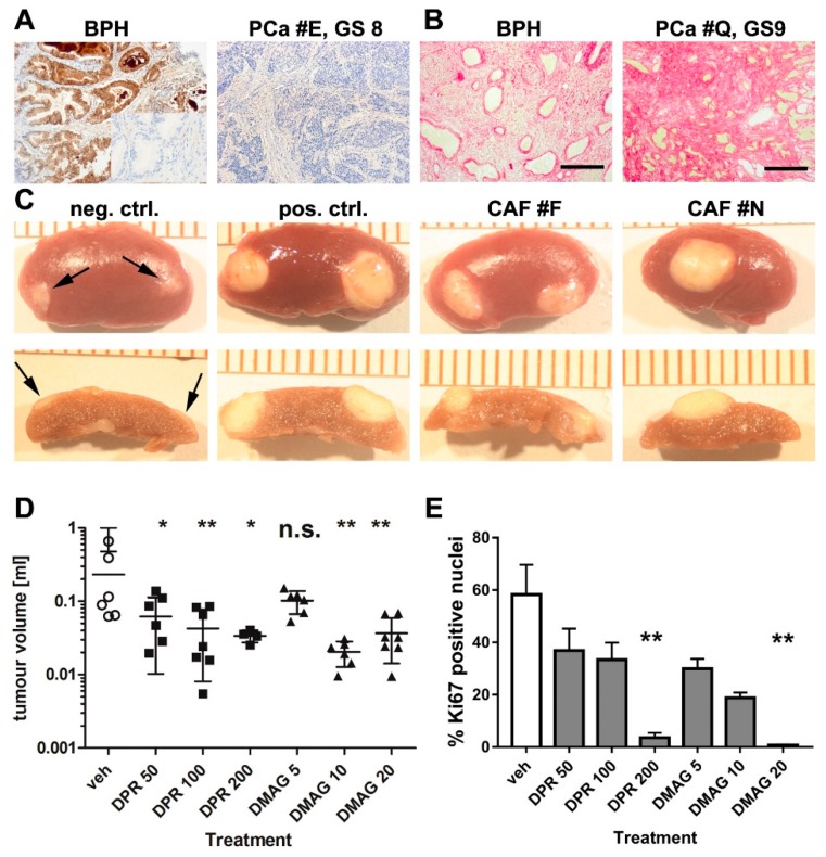
Characterisation of cancer-associated fibroblasts (CAF) and inhibition of tumour size and proliferation after treatment with HSP90 inhibitors. (A) Immunohistochemistry for Microseminoprotein (MSMB) showed positive staining in non-malignant benign prostatic hyperplasia BPH tissue (left panel; inset: antibody control) but was mostly absent in PCa (right panel) and was used to confirm tumour presence in tissue from which CAF were derived. Microscopy with 10× objective lense; PCa panel has identical scale as images in (B) (scale bar 200 µM). BPH images also with 10× objective lense but illustration at half the size as the other images in (A,B); (B) Collagen was stained by picrosirius red in BPH (non-malignant) and prostate cancer with a Gleason score of 9. Collagen was more abundant in prostate cancer samples. Microscopy with 10× objective; scale bar 200 µM; (C) Recombination of normal prostate fibroblasts (neg. ctrl., negative control) with BPH1 cells and kidney capsule grafting produced little growth (arrows). Recombination of previously validated CAF (pos. ctrl., positive control) and CAF isolates F and N with BPH1 cells led to tumour growth, demonstrating pro-tumourigenic activity of our primary CAF. Images in the upper row shows gross morphology, while the lower row shows cross sections of tumours used for size measurements; (D) CAF/BPH1 tumours were grown for 2 months followed by a 1 month treatment with vehicle control (veh), dipalmitoyl-radicicol (DPR) at 50, 100 and 200 mg/kg or with 17-DMAG (DMAG) at 5, 10 and 20 mg/kg. Tumour size was reduced after treatment with dipalmitoyl-radicicol at all doses, and with DMAG at 10 or 20 mg/kg. The vehicle control group was the same for both treatment groups since tests were performed simultaneous in parallel. Data are presented as mean + S.E.M. and asterisks denote the level of significance * for p < 0.05, ** for p < 0.01 and *** for p < 0.001. n.s. = not significant. Statistical test: one-way ANOVA. Individual data are provided in Supplementary Table S2; (E) To examine effects upon cell proliferation within tumours, tissue sections of CAF/BPH1 tumours were stained for the proliferation marker Ki67, and positive nuclei counted. The highest doses of DPR treatment (200 mg/kg) and DMAG treatment (20 mg/kg) resulted in a significantly decreased Ki67 index. n = 3; one way-ANOVA, data are presented as mean + S.E.M. and asterisks denote the level of significance as mentioned above.

