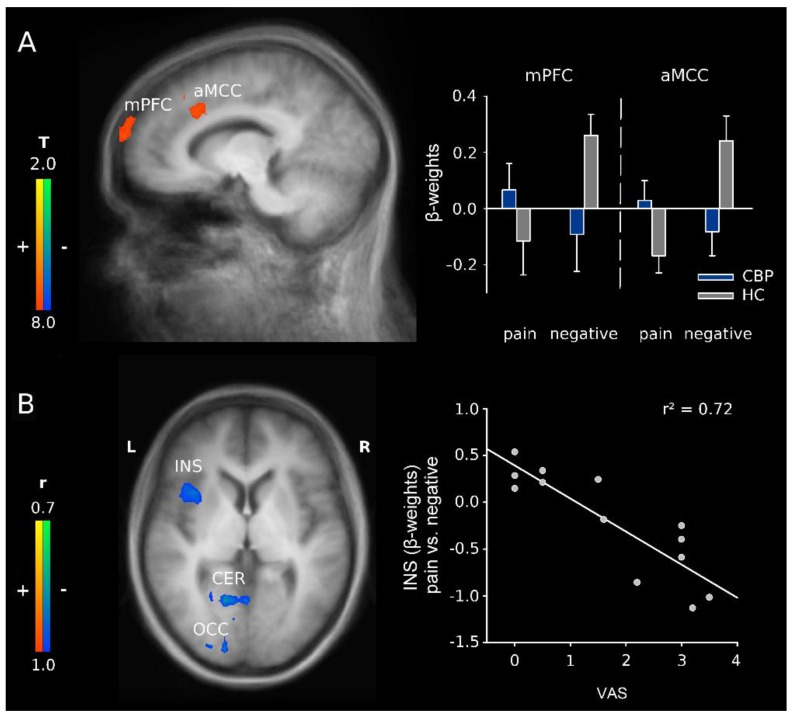
Figure 3.
(A) activation maps illustrating the interaction between group (CBP patients vs. HC) and word category (pain-related vs. negative adjectives) with activations in the medial prefrontal cortex (mPFC) and anterior midcingular cortex (aMCC) including the dorsolateral prefrontal cortex (DLPFC); x = −10. Right: schematic overview of the β-weights for the aforementioned structures; mean + Standard Error; and (B) correlation of current pain (VAS) with the differences in parameter estimates for the contrast pain-related vs. negative adjectives in CBP patients in insula (INS), cerebellum (CER) and occipital cortex (OCC); z = 4. Activations are superimposed on a Talairach template (average of all subjects). Right: correlation plot for the relation of current pain (VAS) and differences in parameter estimates for the contrast pain-related vs. negative adjectives for the anterior insula.

