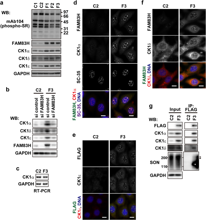
Figure 5. The expression and subcellular localization of CK1α, δ, and ε are regulated by FAM83H.
(a) RKO cells that are stably transfected with the FAM83H-FLAG vector (F1, 2, and 3) or the empty vector (C1 and 2) were analyzed by Western blotting. The numbers at the right side of the top panel indicate the positions of molecular weight markers (kDa). (b) RKO-C1 and F3 cells were transfected with siRNA for FAM83H or control siRNA and then analyzed by Western blotting. (c) RKO-C1 and F3 cells were analyzed for the expression of the indicated mRNA by RT-PCR. (d–f) RKO-C1 and F3 cells were stained with the indicated antibodies and DAPI (for DNA, blue). Scale bars, 10 μm. (g) Immunoprecipitates using an anti-FLAG antibody were prepared from RKO-C1 and F3 cells. Input lysates and immunoprecipitates were analyzed by Western blotting. Arrowheads indicate the migrating positions of major fractions of SON. An asterisk indicates the migrating position of FAM83H-FLAG.

