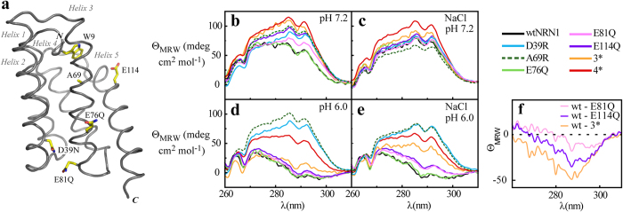
Figure 1. Structural analysis of wildtype and variants of NRN1L.h.
(a) The lowest energy structure of the NRN1L.h. variant 3* was solved by NMR spectroscopy and is used as a structural template to show the position of the individually mutated amino acid residues (PDB accession code: 2N3E). (b–e) Near-UV CD spectra of wtNRN1L.h. and variants thereof at (b) pH 7.2, (c) pH 7.2 in the presence of 300 mM NaCl, (d) pH 6.0, (e) pH 6.0 in the presence of 300 mM NaCl. (f) The near-UV CD spectra of E81Q, E114Q and 3* were subtracted by the spectrum of wtNRN1L.h.to illustrate the significant spectral differences at pH 7.2. All spectra were taken at 142.5 μM.

