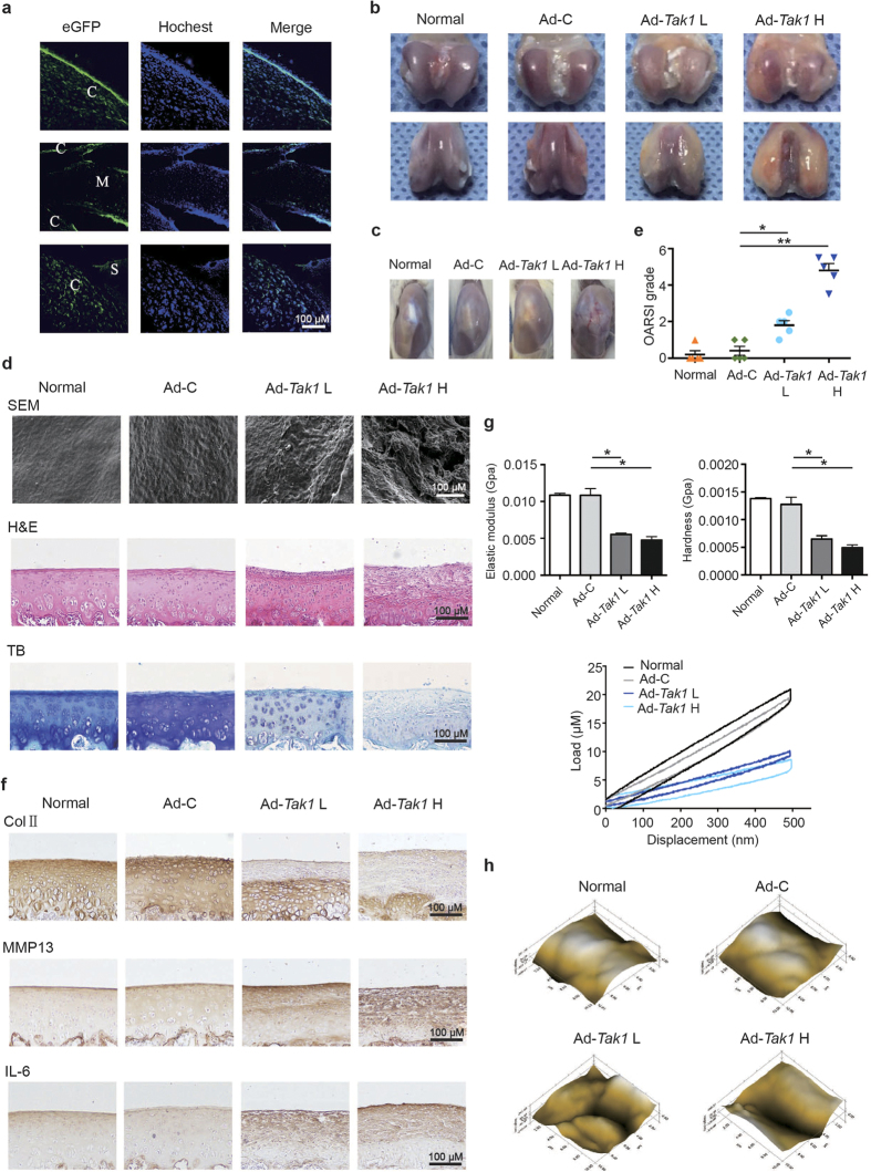
Figure 1. Intra-articular overexpression of TAK1 causes cartilage destruction in rats.
(a) Representative immunofluorescence image of cartilage sections from rats intra-articularly injected with Ad-Tak1. Green: GFP, blue: DAPI. C: cartilage, M: meniscus, S: synovium. Scale bar, 100 μm. (b) Representative images of rat articular cartilage from normal, Ad-C, Ad-Tak1 L and Ad-Tak1 H groups. (c) Representative gross appearance of rat joints from each group. (d) SEM, H&E and toluidine blue staining of articular cartilage from each group. Scale bar, 100 μm. (e) OARSI scores of articular cartilage from each group. *P < 0.05, **P < 0.01. (f) IHC staining of type II collagen, MMP13 and IL-6 of articular cartilage from each group. Scale bar, 100 μm. (g) The biomechanical properties including elastic modulus, hardness and load-displacement curves of cartilage surface from each group. *P < 0.05. (h) Microscopic geomorphology of cartilage surface from each group.

