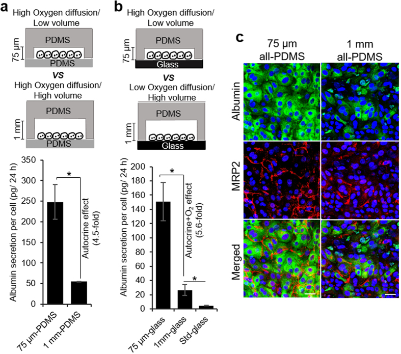
Figure 3. Investigating the effects of oxygen and microchamber volume on phenotype of hepatocytes.
Secretion of albumin by hepatocytes cultured on collagen I-coated PDMS substrate (a) and glass slides (b) in 75 μm and 1 mm tall μCs. Hepatocytes grown for 7 days on collagen I-coated glass slides in 12-well plates were used as control (denoted as standard). The data indicate means ± SD (n = 3). *p < 0.05. (c) Immunostaining images of albumin (green) and MRP2 (red) in hepatocytes cultured for 7 days in all-PDMS microchambers with variable heights. Nuclei are stained by DAPI (blue). Scale bar = 50 μm.

