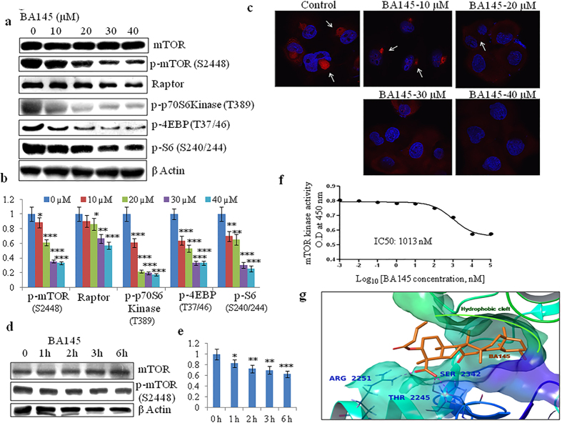
Figure 5. BA145 inhibits mTOR pathway in PANC-1 cells.
(a) PANC-1 cells were treated with BA145 at indicated concentrations for 12 h. Whole cell protein lysates were prepared and the expression levels of mTOR and its downstream substrates were analyzed by western blotting (b) Densitometric analysis of p-mTOR (S2448), Raptor, p-p70S6Kinase (T389), p-4EBP (T37/46) and p-S6(S240/244) protein expression in BA145 treated PANC-1 cells (c) Detection of p-mTOR (serine 2448) by immunofluorescent microscopy in BA145 (12 h) treated PANC-1 cells at different concentrations. BA145 treatment inhibits p-mTOR expression (red arrowheads) in PANC-1 cells in dose dependent manner (d) PANC-1 cells were exposed to BA145 (40 μM) at 1 h, 2 h, 3 h, 4 h and 6 h time intervals and the expression of mTOR and p-mTOR proteins were examined by western blotting. β-actin was used as a loading control (e) Densitometric analysis of p-mTOR expression (f) BA145 inhibits mTOR kinase in cell free mTOR kinase enzyme assay (g) 3D Interaction diagram of BA145 (in orange) with mTOR. The fused rings of the ligand are enclosed by the hydrophobic cleft formed within the binding pocket and the residues forming H-bond with the carboxylic group of the ligand are also shown in bold. The glide score of the complex was found to be −10.49 Kcal/mol. Columns, mean; bars, SD with ***p < 0.001, **p < 0.01, *p < 0.05 versus control.

