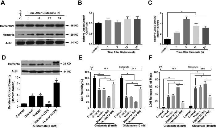
Figure 1. Increased Homer1a protein levels improved HT-22 cell survival after glutamate treatment.
Twenty-four h after seeding the cells into 60-mm plates, the HT-22 cells were exposed to glutamate (10 mM) for either 1, 6, 12 or 24 h. The proteins levels of Homer1b/c and Homer1a were evaluated by western blotting (A). The relative expressions of Homer1b/c (B), and Homer1a (C) were calculated as percent of the optical density of the control group. After infection with either Homer1a shRNA or Homer1a O/E lentiviral vectors, the HT-22 cells were exposed to glutamate (5 mM) for 6 h, after which the proteins were harvested for western blotting assay (D). Forty-eight h after lentiviral infection, followed by 24 h of glutamate treatment, cellular viability was assessed by CCK-8 assay as a percentage of control group (E), and the cell injury was detected by LDH release assay (F). HT-22 cells in the control group were not subjected to lentiviral infection or glutamate injury. Data are presented as mean ± SEM from four experiments; *p < 0.05. #p < 0.05 vs. control + glutamate or vector + glutamate group.

