
Figure 5. ERI3 localizes to Golgi structures and COPII vesicles.
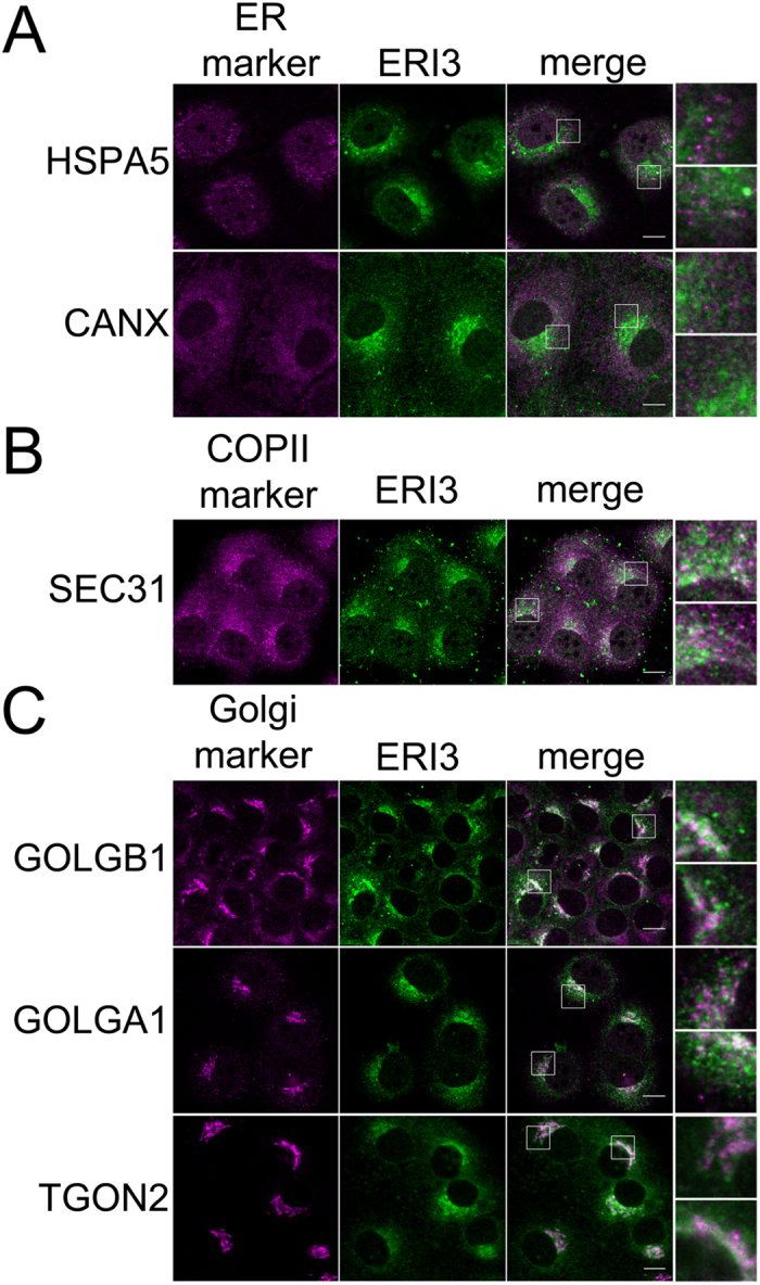
HuH-7 cells were plated on coverslips and fixed, permeabilized and probed with antibody to ERI3 and the indicated organellar marker protein. Fluorescent confocal micrographs show (from left to right) organellar marker, ERI3, overlay, and insets from the indicated region in the overlay panel. (A) Localization of ERI3 and the ER markers HSPA5 and CANX. (B) Localization of ERI3 and the COPII marker Sec31. (C) Localization of ERI3 and the Golgi markers GOLGB1, GOLGA1 and TGON2. Scale bar = 10 μm. Localization was analyzed in at least three independent experiments.