
Figure 6. ERI3 relocalizes to viral replication sites in DENV-2-infected cells.
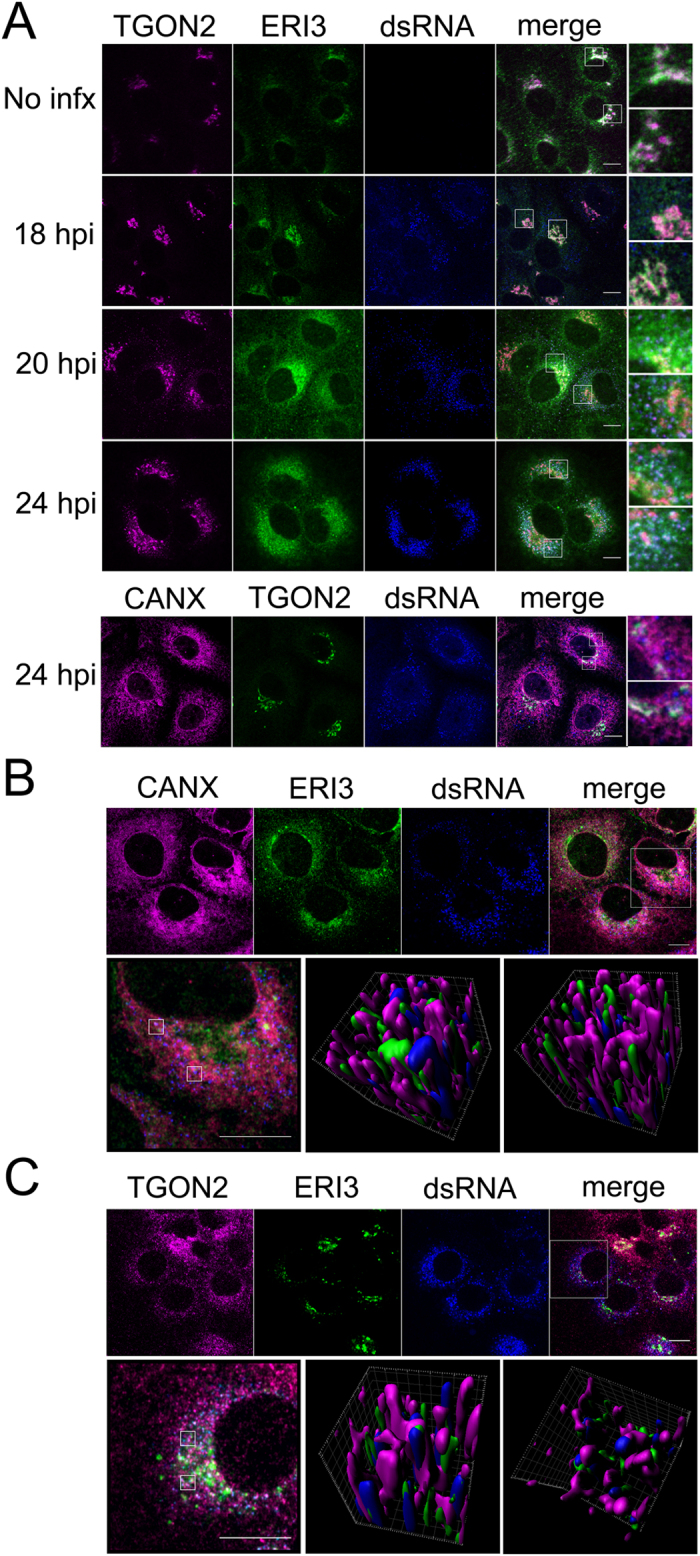
HuH-7 cells were plated on coverslips and infected with DENV-2. At the indicated time post-infection, cells were fixed, permeabilized and probed with antibodies. Fluorescent confocal micrographs show (from left to right) ERI3, dsRNA, the indicated organellar marker, overlay, and insets from the indicated region in the overlay panel. (A) Coverslips from different timepoints post-infection were probed with antibodies to ERI3, dsRNA and either the Golgi marker TGON2 or the ER marker CANX. (B,C) Coverslips from 24 hour DENV-2 infections were probed with antibodies to ERI3, dsRNA and the ER marker CANX or Golgi marker TGON2. Three-dimensional rendering of the regions identified in the insets were generated using the Imaris software package. Localization was analyzed in at least three independent experiments.