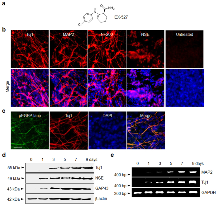
Figure 1. EX-527 promotes neuronal differentiation in P19 cells.
(a) Chemical structure of EX-527. (b) P19 cells were incubated with 100 μM EX-527 for 9 days. (Upper panel) The treated cells were immunostained with antibodies against neuron-specific proteins. (Lower panel) Merged images of cells treated with antibodies and DAPI (blue). ‘Untreated’ indicates no treatment of P19 cells with EX-527. Scale bar, 50 μm. (c) P19 cells stably transfected with a Tau promoter-EGFP fusion gene (pEGFP-taup) were incubated with 100 μM EX-527 for 9 days. The treated cells were immunostained with anti-Tuj1 antibody (scale bar; 50 μm). EGFP (green) and Tuj1 (red) colocalize in the treated P19 cells. (d) The expression levels of neuron-specific markers in P19 cells treated with 100 μM EX-527 were examined at various times by using western blot and (e) RT-PCR analyses. β-Actin and GAPDH were used as loading controls.

